http://www.collembola.org/publicat/morpholo/fat.htm
-
Last updated on
2024.08.02
by Frans Janssens
Frans Janssens,
Department of Biology, University of Antwerp, Antwerp, B-2020, Belgium
Abstract.
To be completed.
Introduction
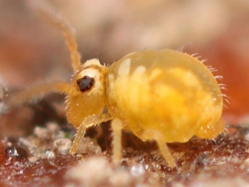
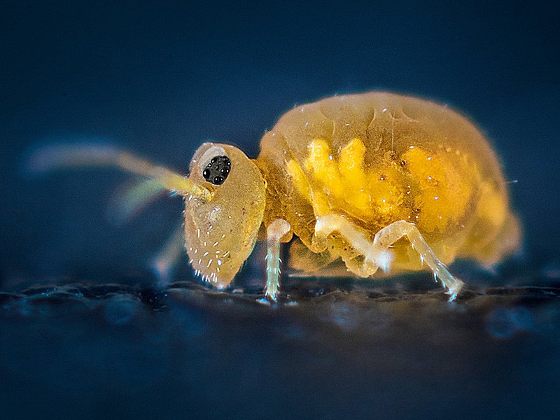
Fig.Sa. Sminthurinus aureus from Belgium
2021.01.19 © Huskens, M.L.
 |
The subcutaneous fat tissue may affect the appearance of the dorso-lateral
colouration in habitus images of unpigmented or poorly pigmented specimens
(fig.Sa).
To be completed.
Case studies
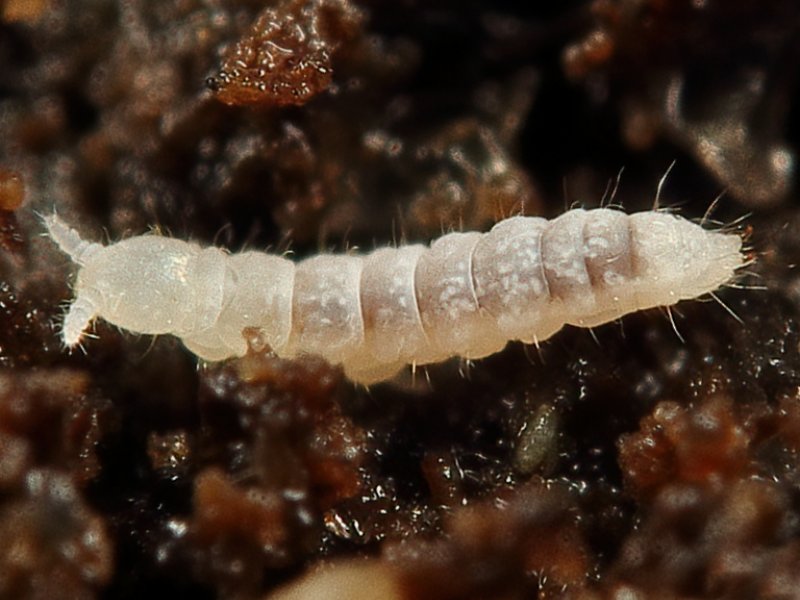
Neanuridae: Sensillanura barberi
Fig.Sb. Sensillanura barberi from the USA
2024.06.02 © Coogler, J.
 |
In Sensillanura barberi,
the fat tissue is distributed in small globulae
and appears as dorso-lateral white
spots, scattered on head, thorax and abdomen
(fig.Sb).
To be completed.
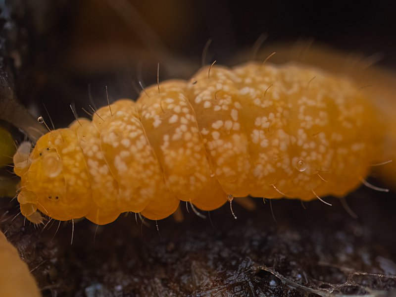
Onychiuridae: Onychiurinae
Fig.Osp. Onychiurinae from Canada
2024.07.30 © Chan, J.
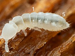 |
In Onychiurinae,
the fat tissue appears as dorso-lateral white
spots, present on abdominal segments 1 to 5, absent on abdominal segment 6.
They are especially distinctly visible in the feeding instar,
when the midgut is filled with dark ingested food,
and the food serves as dark background (fig.Osp).
To be completed.
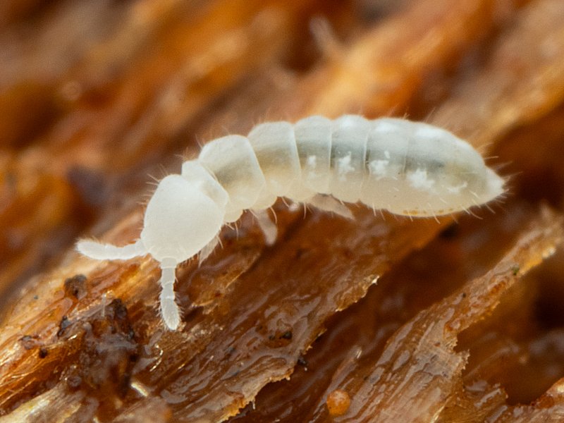
Tullbergiidae: Paratullbergia callipygos
Fig.Pc. Paratullbergia callipygos from Holland
2024.04.02 © Kamsteeg, G.
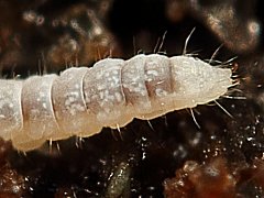 |
In Paratullbergia callipygos,
the fat tissue appears as dorso-lateral white
spots, scattered on thorax and abdomen,
and especially distinctly visible in the feeding instar,
when the midgut is filled with dark ingested food,
and the food serves as dark background (fig.Pc).
To be completed.
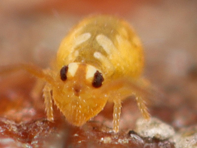
Katiannidae: Sminthurinus aureus
Fig.Sa3. Sminthurinus aureus from Germany
2014.10.20 © Gutekunst, V.
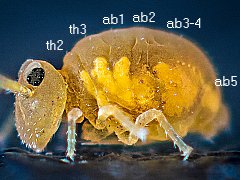 |
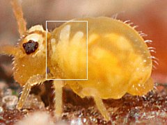
Fig.Sa2. Sminthurinus aureus from Belgium
2021.01.19 © Huskens, M.L.
 |
In Sminthurinus aureus,
the fat tissue appears as dorso-lateral yellow bands or spots
on the thoracic and abdominal segments
(fig.Sa, fig.Sa2, fig.Sa3).
The fat tissue is organised per segment and marks as such the bounderies
of the thoracic and abdominal segments in the great abdomen (fig.Sa3).
In the anterior part of the great abdomen, the fat tissue
forms yellow bands that radiate from the ventrum to the dorsum.
The bands are distinctly visible in the 2nd and 3rd thoracic segments
and in the 1st abdominal segment.
In the 2nd to 4th abdominal segments,
the fat tissue is visible as a more or less fused lateral yellow spot,
while in the 5th abdominal segment it is a clearly separated spot (fig.Sa3).
The fat tissue forms as such a lateral band that circumvents the large abdomen.
In addition, notice the darker dorsum, which is typical for a feeding instar
in which the gut is filled with ingested food, improving as such the contrast
with the fat tissue.
To be completed.
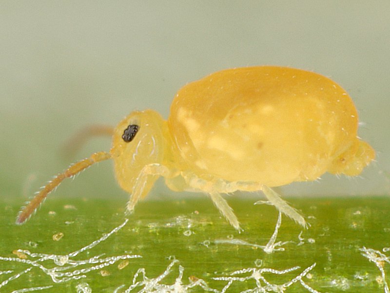
Bourletiellidae: Deuterosminthurus pallipes repandus
Fig.Dpr. Deuterosminthurus pallipes repandus ♀ from Belgium
2024.04.25 © Huskens, M.L.
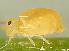 |
In Deuterosminthurus pallipes repandus,
the fat tissue appears as a dorso-lateral yellow transverse band
on the 3rd thoracic segment
(fig.Dpr).
Notice the darker dorsum, which is typical for a feeding instar
in which the gut is filled with ingested food, improving as such the contrast
with the fat tissue.
To be completed.
Urate crystals embedded in fat tissue
In the fat tissue, urate crystals may be deposited. In such case the fat tissue
seems to glow due to reflection of the flash light of the illumination
in the crystals.
To be completed.
Discussion
To be completed.
Conclusion
To be completed.
Acknowledgements
We would like to thank, in alphabetical order,
Justin Chan,
Joshua Coogler,
Valentin Gutekunst,
Marie Louise Huskens,
Gertjan Kamsteeg,
for their kind permission for using their respective photographs
as illustrations.
References