http://www.collembola.org/publicat/morpholo/gut.htm
-
Last updated on
2024.09.03
by Frans Janssens
Frans Janssens,
Department of Biology, University of Antwerp, Antwerp, B-2020, Belgium
Abstract.
Skin pigmentation has been a neglected diagnostic character given it is quite
variable in instars of the same species. But also due to complexities caused
by metabolic issues such as ingestion of food.
Here we will explore the effect of the colour of the ingested food in the gut
on the appearance of the habitus colouration.
To be completed.
Introduction
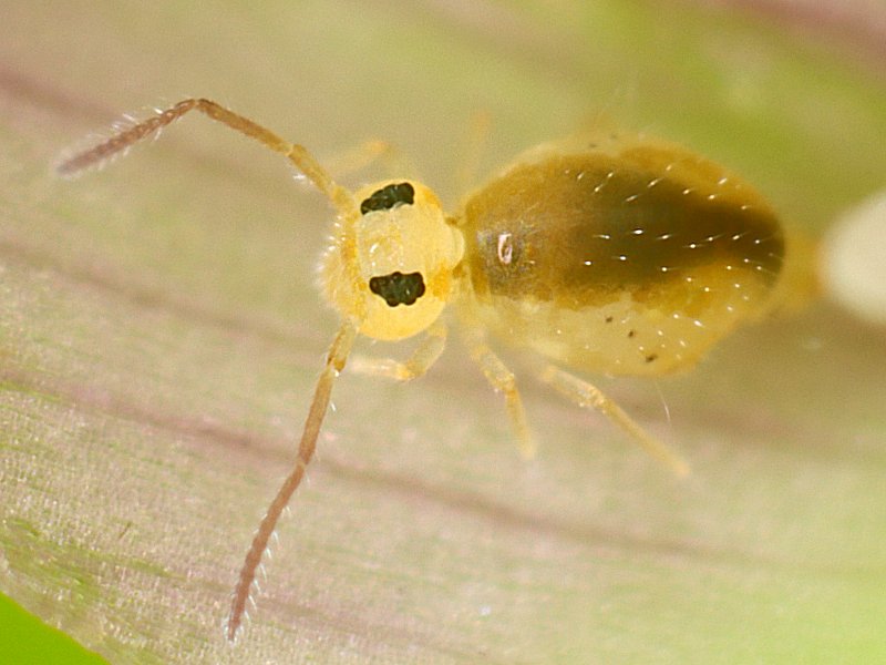
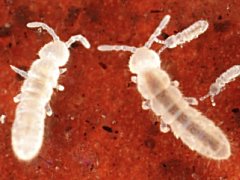
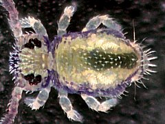
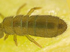
Fig.Do1. Dicyrtomina ornata juv. from the UK
2024.02.22 © Barton, T.
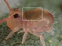 |
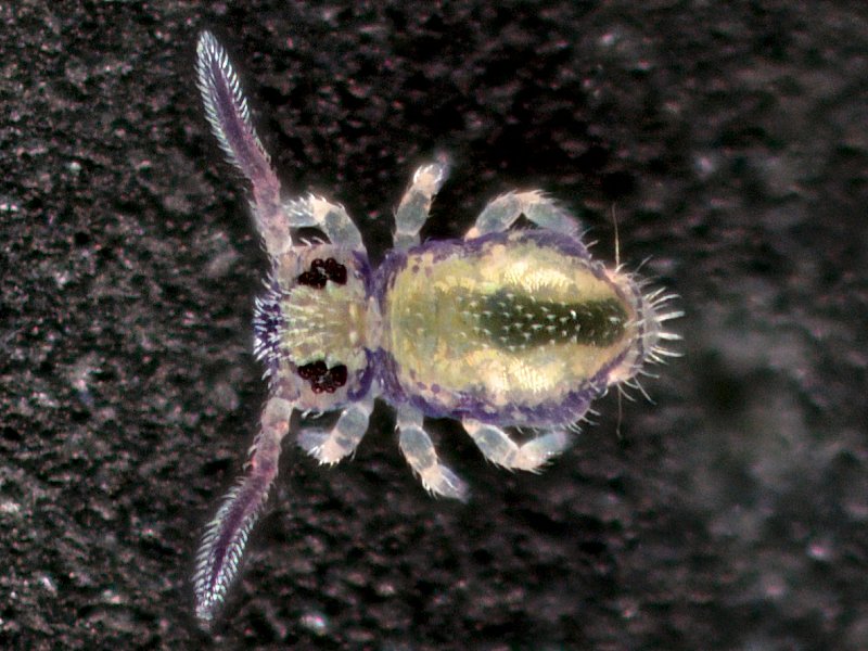
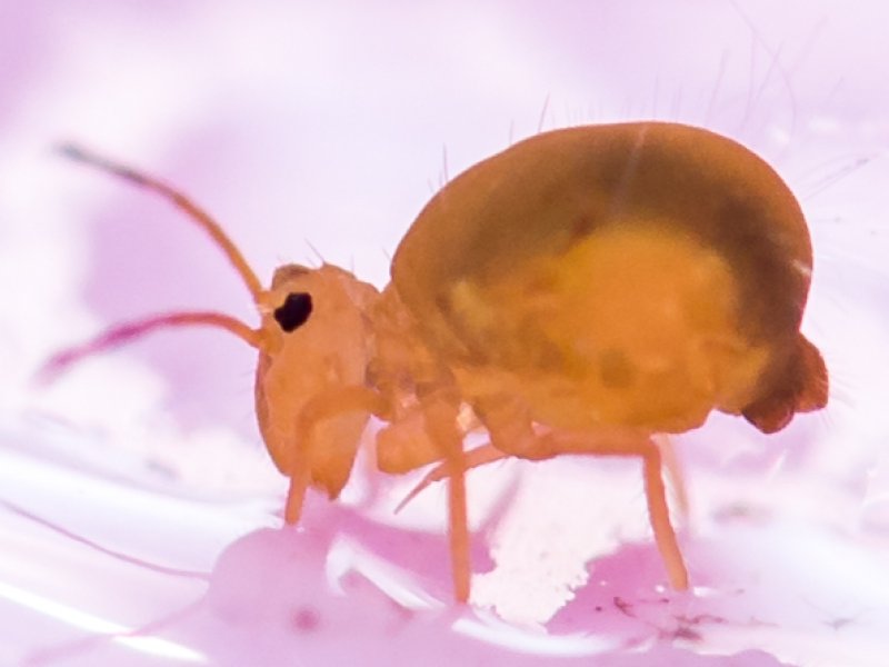
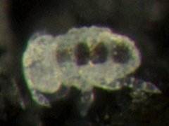
Fig.NMm2. Megalothorax minimus from Belgium
2020.02.05 © Huskens, M.-L.
 |
The gut of the feeding instar of Collembola is typically filled with ingested
food. The colour of the ingested food may affect the appearance of the dorsal
colouration in habitus images of unpigmented or poorly pigmented specimens
(fig.Do1, fig.NMm2).
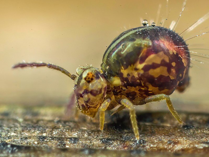
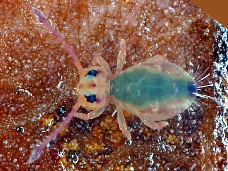
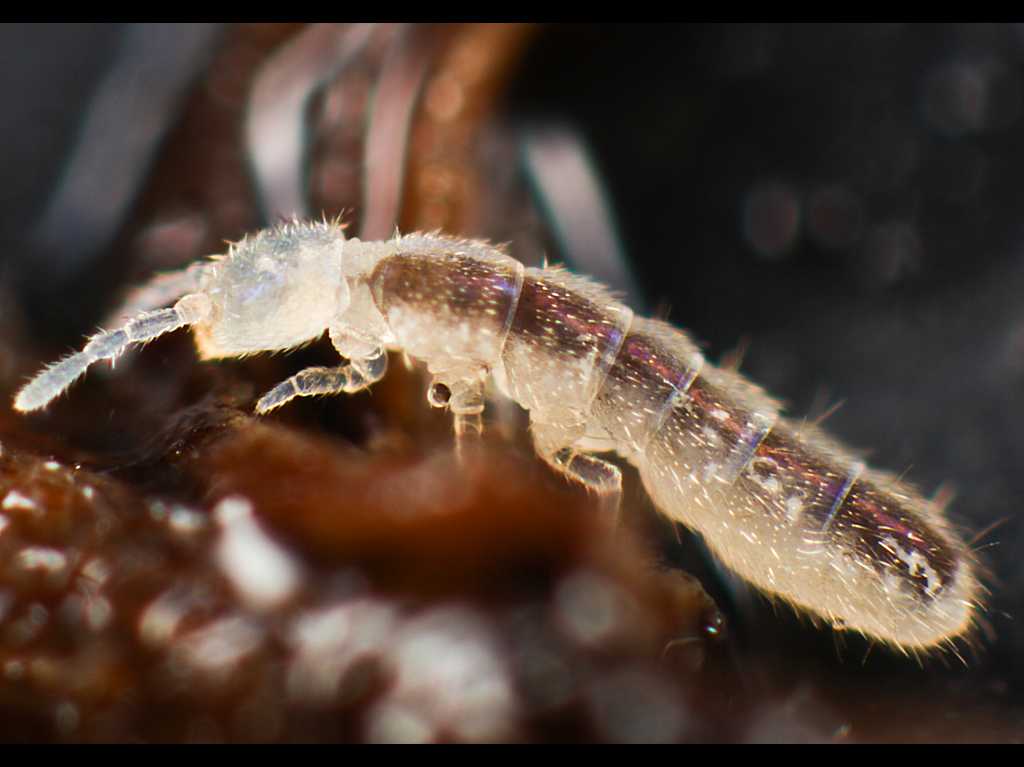
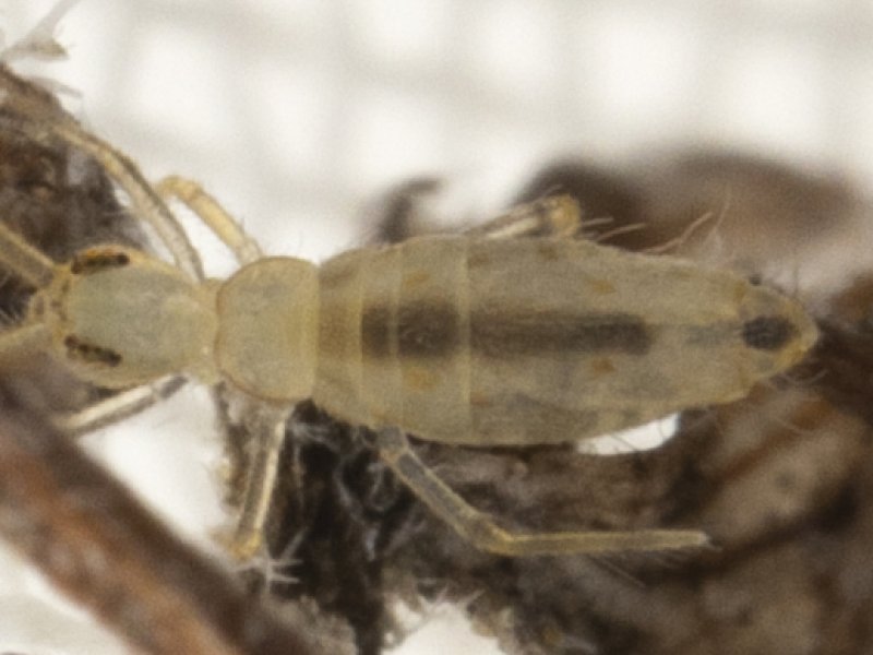
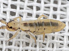
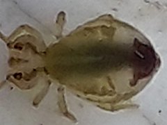
Fig.Jnrs. Jordanathrix near superba from the UK
2024.02.15 © Phillips, E.
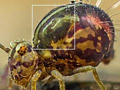 |
Fig.Do2. Dicyrtomina ornata from the UK
2024.02.22 © Barton, T.
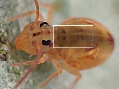 |
The filled gut may obfuscate the intrinsic body pigmentation
in patterned specimens
(fig.Jnrs, fig.Do2).
In addition the intrinsic shape of the gut may also
influence the appearance of the dorsal colouration (fig.NMm).
To be completed.
Digestive system
The digestive system of Collembola consists of
a narrow tubular foregut, running from the mouth to the first thoracic segment,
an enlarged midgut, running in the second thoracic segment to the fourth abdominal segment,
a narrow tubular hindgut, running in the fifth abdominal segment to the
6th abdominal segment,
and the expanded rectum in the 6th abdominal segment that opens at the anus
(Betsch, 1980:Fig.4).
The foregut, hindgut and rectum are lined with a thin cuticula
which is shed at every moult.
(modified after Hopkin 1997:60).
In the midgut epithelium waste material is accumulated in granules.
Towards the end of the intermoult period, the epithelium degenerates
and is released in the midgut lumen just before ecdysis
and a new epithelium is formed.
(modified after Hopkin 1997:63-64).
To be completed.
Reproductive instar versus feeding instar
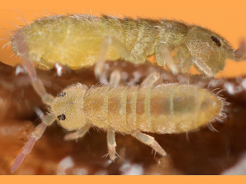
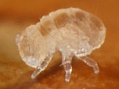
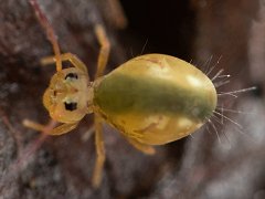
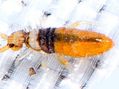
Fig.Fc. Folsomia candida from Germany
Left reproductive instar, right feeding instar
2006.12.26 © Kohl, F.
 |
Contrary to Insecta, adult Collembola keep on moulting.
During the adult phase two distinct types of instars occur.
The reproductive instar in which the gut is empty (fig.Fc left).
The feeding instar in which the gut is filled with ingested food (fig.Fc right).
Only in the reproductive instar the intrinsic colouration of the species
can be observed.
In the feeding instar the colouration may be obfuscated by the colour of the
food in the midgut.
The dorsal median longitudinal brown stripe observed in the feeding instar
of Folsomia candida is not skin pigmentation but it is the colour of the
ingested food in the midgut as seen through the translucent body.
To be completed.
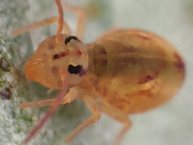
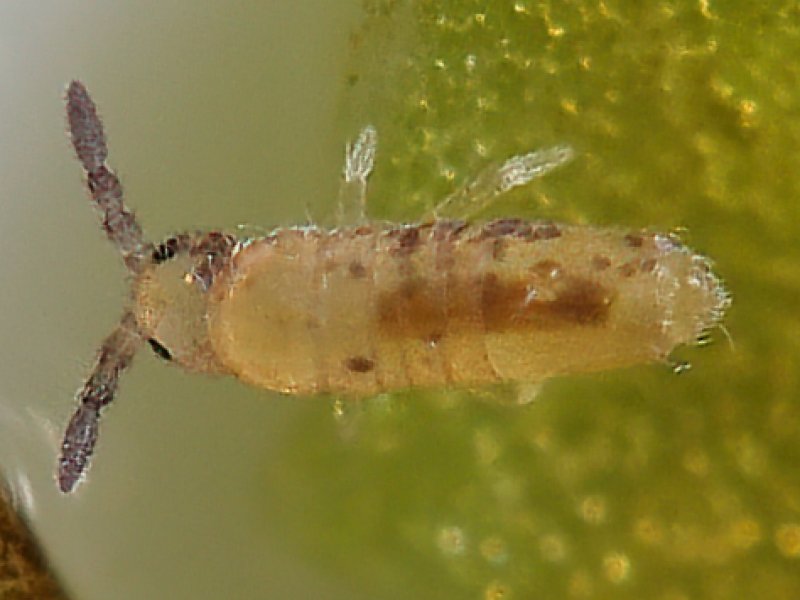
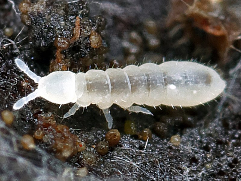
Juvenile instars
Fig.SS. Sminthurus sp. juv. from the USA
2020.07.12 © Cam, C.
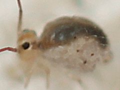 |
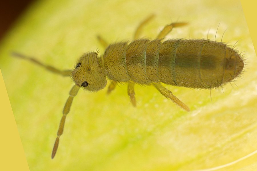
Fig.Oa. Orchesella alpa juv. from the USA
2024.01.06 © Hendrix, S.
 |
Juvenile instars are also feeding instars
in which the midgut is always filled with ingested food
(fig.SS, fig.Oa, fig.Sb2, fig.Hb).
Fig.EL. Lepidocyrtus sp. juv. from Holland
Descaled
2024.02.23 © Kamsteeg, G.
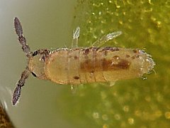 |
Fig.Ps. Pseudobourletiella spinata juv. from the USA
Hatchling
2024.06.20 © Gruver, B.
 |
The midgut can also be partly filled (fig.EL,fig.Ps).
To be completed.
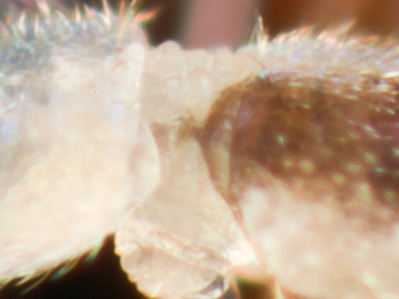
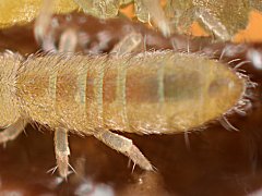
Narrow tubular foregut
Fig.Fcfg. Folsomia candida from the UK
Narrow foregut
2013.01.26 © Murray, A.
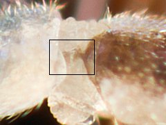 |
The narrow tubular foregut connects to the distinctly more wide midgut
in the prothorax (fig.Fcfg).
To be completed.
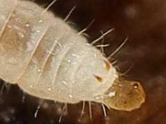
Appearance of filled midgut in the 4 different Collembola orders
Neelipleona
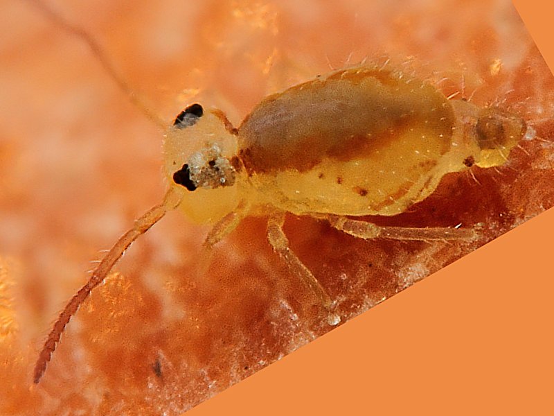
Fig.NMm. Megalothorax minimus from the USA
2011.01.07 © Gerber, G.K.
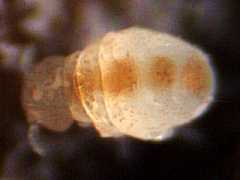 |
Given the diverticulate nature of the midgut in Neelipleona,
the with food filled gut manifests itself as
a dorsal median longitudinal pattern of 4 equally sized coloured discs
(fig.NMm).
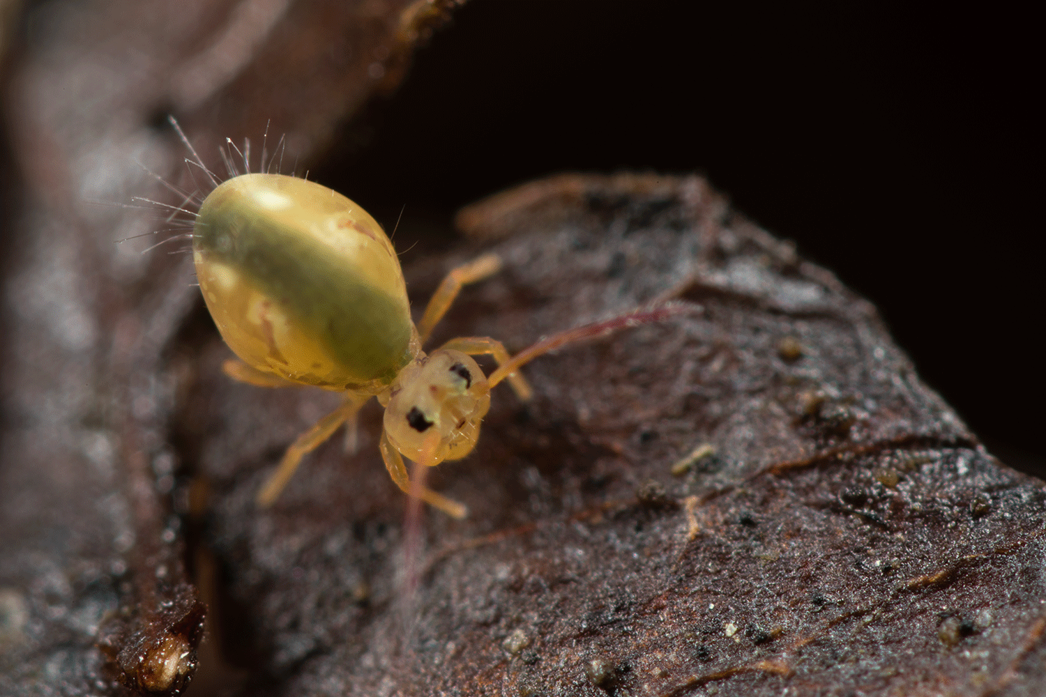
Fig.NMs2. Megalothorax sp. juv. from Germany
2015.01.07 © Mattew, M.
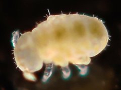 |
Fig.NMs. Megalothorax sp. juv. from Taiwan
2022.03.29 © Cheng, H.-J.
 |
The colour of the discs is variable
and depends on the colour of the ingested food, such as
brown/orange (fig.NMm),
green (fig.NMs2)
or
black (fig.NMs).
Symphypleona
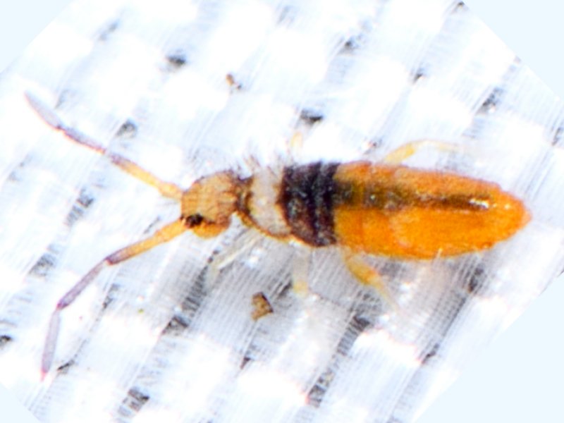
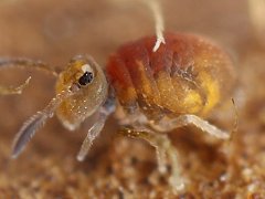
Fig.SHb. Heterosminthurus bilineatus from France
2017.09.03 © Tiky, S.
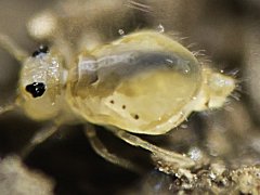 |
Fig.SDo. Dicyrtomina ornata from France
2018.12.04 © Garcelon, P.
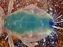 |
Fig.Hb. Heterosminthurus bilineatus from Belgium
2024.08.30 © Huskens,%M.-L.
 |
The anterior part of the midgut is distinctly wider than the posterior part
(fig.SDo, fig.SS, fig.SHb). The colour of the gut contents obfuscates the
intrinsic colouration of the specimen (fig.Hb).
Fig.SDm. Dicyrtomina minuta from Sweden
2016.09.19 © Neergaard, R.
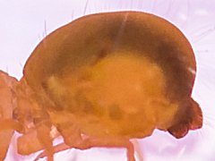 |
Fig.SSd. Sminthurinus domesticus from Belgium
2019.03.13 © Huskens, M.-L.
 |
The midgut is upcurved dorsally (fig.SDm, fig.SS, fig.SSd).
The colour of the ingested food in the anterior part of the midgut
is well visible in the dorsal thoracic region (fig.SSd).
Due to the carotene, it is distinctly reddish in Sminthurinus domesticus,
held in a culture on coocked carots (fig.SSd).

Fig.SJnrs. Jordanathrix nr superba juv. from Belgium
2017.09.29 © Janssens, F.
|
Fig.SDs. Dicyrtomina signata from France
2017.10.07 © Picard, J.
 |
The posterior part of the filled midgut continuously
wiggles left-right in the body cavity (animations fig.SJnrs, fig.SDs).
Poduromorpha
Fig.PO. Onychiurinae from France
2019.10.31 © Garcelon, P.
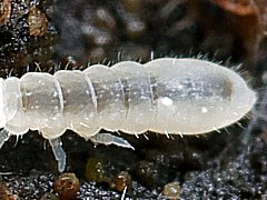 |
Fig.Cd. Ceratophysella denticulata-group from the USA
2024.02.27 © Coogler, J.
 |
The gut is tubular and runs from the mesothorax to the fourth abdominal segment
(fig.PO, fig.Cd).
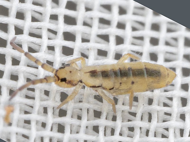
Entomobryomorpha
Fig.EFc. Folsomia candida from the UK
2013.01.26 © Murray, A.
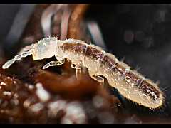 |
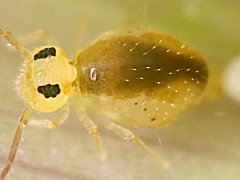
Fig.EEa. Entomobrya atrocincta ♂ from the USA
2021.12.17 © Wildwings.
 |
The gut is tubular and runs from the mesothorax
to the end of the fourth abdominal segment
(fig.EIg, fig.EFc, fig.S, fig.EEa).
When the midgut is partly filled,
it appears shorter,
such as in this descaled juvenile Lepidocyrtus sp. from Holland,
in which it appears to run
from the first to the fourth abdominal segment (fig.EL).
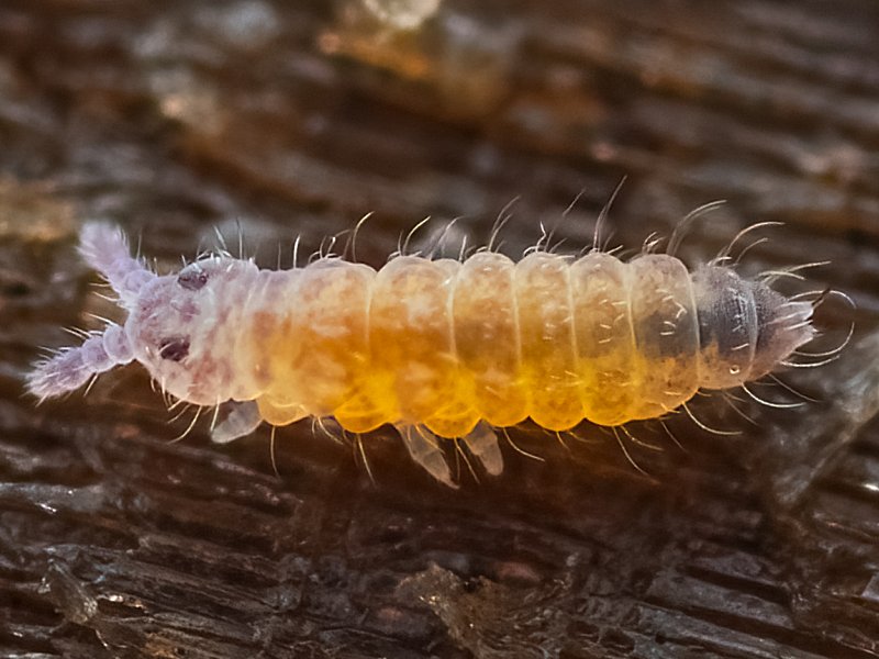
Faecal pellet formation
Fig.Cd2. Ceratophysella denticulata-group from the USA
2024.02.27 © Coogler, J.
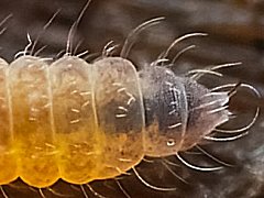 |
Fig.Fq. Folsomia quadrioculata juv. from Holland
2024.03.13 © Kamsteeg, G.
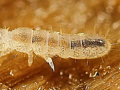 |
Fig.If. Pale colour form of Isotomurus fucicolus juv. from Belgium
2024.08.03 © Huskens, M.-L.
 |
The digested food in the midgut may become darker towards the end of the midgut,
an indication of the precursor of the faecal pellet
(fig.Cd2, fig.Fq, fig.If).
Fig.ECt. Coecobrya tenebricosa juv. from the USA
2017.02.08 © Abbott, J.C.
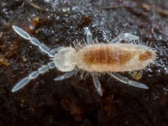 |
Fig.S. Salina sp. juv. from Singapore
2023.10.24 © Budak.
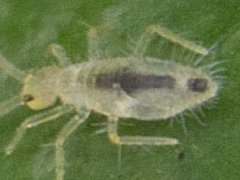 |
Fig.EIg. Isotomurus graminis from Belgium
2024.02.13 © Huskens, M.-L.
 |
Later the posterior part of the midgut contents is split off and transported to
the hindgut. The faecal pellet in the making in the hindgut may affect
the appearance of the colouration of the fourth and fifth abdominal segment.
Then the gutcontents presents itself as a bipartite dorsal median longitudinal
stripe, with a long anterior part of the midgut
and a short posterior part of the hindgut.
(fig.EIg, fig.Cd, fig.S, fig.Sb, fig.Sb2, fig.ECt).
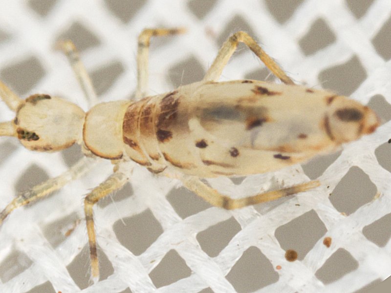
Fig.Sb2. Salina banksi juv. from the USA
2024.01.07 © Hendrix, S.
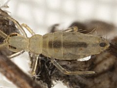 |
Fig.Sb. Salina banksi from the USA
2024.01.12 © Hendrix, S.
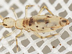 |
Fig.Hb. Heterosminthurus bilineatus juv. from Holland
2024.04.30 © Kamsteeg, G.
 |
In patterned specimens,
the dark colour of the midgut contents and of the faecal pellet in the hindgut
may obfuscate the true pattern of the skin pigmentation pattern
(fig.Sb, fig.Hb).
To be completed.
Fig.Sv. Sminthurus viridis from Holland
2024.04.08 © Huskens, M.-L.
Contrast enhanced
 |
In uniformly coloured specimens,
the dark colour of the faecal pellet in the hindgut may
suggest the presence of a dark pigmentation band at the 5th abdominal segment
(fig.Sv).
The size of the faecal pellet matches the size of the rectum (= posterior
part of the hindgut) in the 5th abdominal segment
(fig.EIg, fig.Sb, fig.Sb2),
In juveniles, the pellet may be larger then the 5th abdominal segment
(fig.Ecr, fig.S).
In Bourletiellidae, the 5th abdominal segment is more prominent,
and the faecal pellet may be shorter (fig.Hb).
In Sminthurus viridis,
the presence of a faecal pellet in the hindgut
marks the anterior delimitation of the genital segment
(fig.Sv).
To be completed.
Faecal pellet deposition
Fig.Pc. Paratullbergia callipygos from Holland
2024.03.27 © Kamsteeg, G.
 |
After each moult, a faecal pellet is formed in the rectum
(= the expanded terminal part of the hindgut).
With the depostion of that faecal pellet,
the originally in the midgut accumulated waste granules
are evacuated
(modified after Hopkin 1997:64).
After deposition of a pellet,
the colouration of the fifth and sixth abdominal segment
are restored to the intrinsic body colour
(fig.Pc).
To be completed.
Discussion
To be completed.
Conclusion
To be completed.
Acknowledgements
We would like to thank, in alphabetical order,
John C. Abbott,
Toby Barton,
Budak,
Caleb Cam,
Hsin-Ju Cheng,
Philippe Garcelon,
G.K. Gerber,
Ben Gruver,
Solomon Hendrix,
Marie Louise Huskens,
Gertjan Kamsteeg,
Frithjof Kohl,
Mat Mattew,
Andy Murray,
Raimo Neergaard,
Ed Phillips,
Jérôme Picard,
Sun Ticky,
and
Wildwings
for their kind permission for using their respective photographs
as illustrations.
References