http://www.collembola.org/publicat/morpholo/ocellus.htm
-
Last updated on
2024.08.02
by Frans Janssens
Frans Janssens,
Department of Biology, University of Antwerp, Antwerp, B-2020, Belgium
Abstract.
To be completed.
Introduction
Fig.Ps1. Ptenothrix sp. from Canada
Outer lateral ocelli transparent
2011.10.26 © Hochhalter, R.
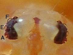 |
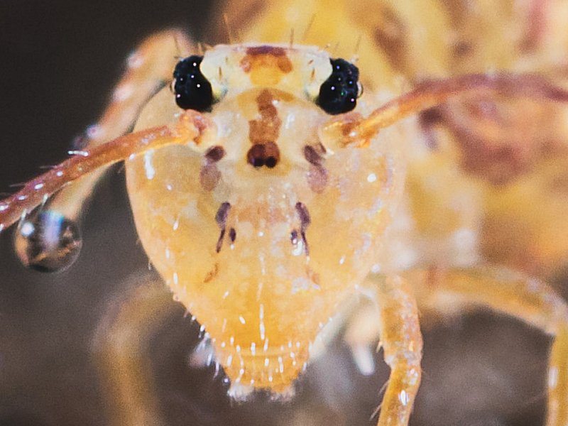
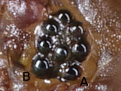
Fig.Md. Morulina delicata juv. from the USA
Left eye
3 anterior + 2 posterior ocelli
2024.06.02 © Coogler, J.
 |
The ocelli of the eye appear differently in frontal and lateral aspect.
In frontal aspect the ocellus appears as a black sphere,
while in lateral aspect it appears as a transparent dome.
In frontal aspect of the eye of Ptenothrix sp.,
the outer lateral ocelli A, E and F appear as transparent domes
(fig.Ps1).
In the eye of the juvenile Morulina delicata,
the 3 anterior ocelli appear as black spheres
while the 2 posterior ocelli appear as transparent domes,
which could be easily overlooked
(fig.Md).
This difference in appearance may complicate counting
and observing the ocelli of the eye.
To be completed.
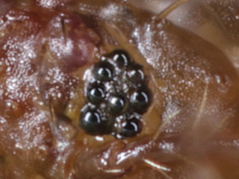
Ocellus observed in frontal aspect
When observed in frontal aspect, the sphere shaped ocellus appears dark.
It is due to the apical structural optical component of the ocellus :
a crystalline transparent sphere focusses the incoming light on
the dark pigment cells of the ocellus?
(fig.Md).
To be completed.
Ocellus observed in lateral aspect
When observed in lateral aspect, the dome shaped ocellus may appear transparent.
That is due to the structural optical component of the ocellus :
the apical crystalline transparent sphere.
In lateral aspect the dark pigment cannot be observed, given
the crystalline sphere does not focus the incoming light on the pigment cells
of the ocellus.
To be completed. �
Heterosminthurus bilineatus
Fig.Hbi. Heterosminthurus bilineatus ♂ from Belgium
Inner dorsal ocelli transparent
2024.07.28 © Huskens, M.-L.
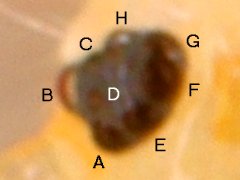 |
In Heterosminthurus bilineatus,
ocelli B, H and G (the inner dorsal ocelli)
may appear transparent (fig.Hbi).
Notice the weird image formed in the transparent ocellus G.
Notice the large prominent size of ocellus B,
an adapation to the peculiar courtship behaviour of this species.
To be completed. �
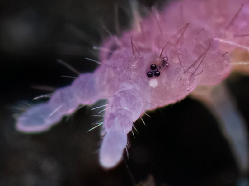
Ptenothrix sp.
Fig.Ps1. Ptenothrix sp. from Canada
Outer lateral ocelli transparent
2011.10.26 © Hochhalter, R.
|
In Ptenothrix sp.,
ocelli A, E and F (the outer lateral ocelli)
may appear transparent (fig.Ps1).
Notice the weird image formed in the transparent ocellus A.
To be completed. �
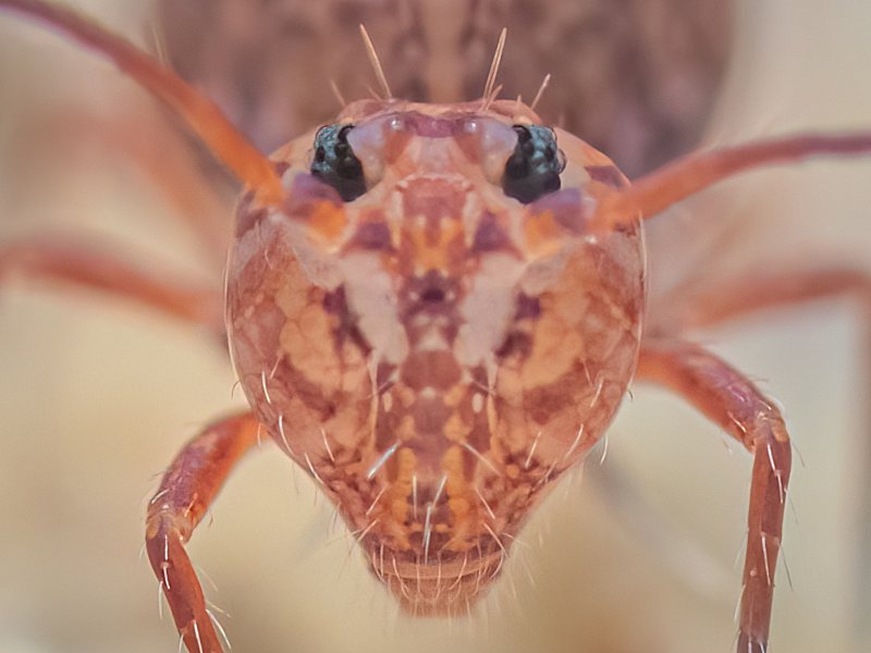
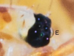
Ptenothrix marmorata f. testudineata
Fig.Pmt. Ptenothrix marmorata f. testudineata from the USA
Left eye in frontal aspect
Ocellus E in lateral aspect
2024.06.15 © Coogler, J.
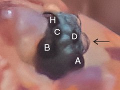 |
Fig.Pmt2. Ptenothrix marmorata f. testudineata from the USA
Left eye in frontal aspect
Ocellus E in lateral aspect in close-up
2024.06.15 © Coogler, J.
 |
In Ptenothrix marmorata f. testudineata,
at least ocellus E,
may appear transparent
(fig.Pmt, fig.Pmt2).
Apically, ocellus E seems to have a kind of lens shaped nipple (fig.Pmt2).
The transparent dome is about 74% high compared to the radius of the ocellus.
To be completed.
Highlights
Depending on the kind of illumination,
one or more highlights may be seen in the ocellus.
With diffuse illumination the highlight is absent.
With bilateral illumination 2 highlights may be present.
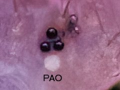
Fig.Dmn. Dicyrtomina minuta f. nigromaculata from France
Left eye in frontal aspect
Ocellus E in lateral aspect
2020.11.18 © Fiacre, R.
 |
A single highlight appears if one lightsource has been used
(fig.Dmn).
To be completed.
Fig.Af. Allacma fusca from Belgium
Left eye in orthogonal aspect
2017.07.16 © Huskens, M.-L.
 |
Duplicate highlights appear if bilateral lightsources have been used
(fig.Af).
Bilateral illumination clearly complicates
the interpretation of the image of the composed eye.
Notice that each ocellus seems to be divided in two parts :
a ventrad gray part
and a dorsad black part in which the highlights are visible.
To be completed.
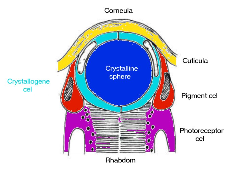
Anatomy of the ocellus
Fig.Ana. Collembola ocellus simplified model
Axial section
2024.06.21 © Janssens, F.
Modified after Paulus, H.F. in Horridge, G.A. 1974 Fig.1.10.f
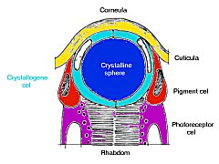 |
The ocellus is a product of the intugement
and is formed by specialised epidermal cells.
It is apically topped by transparent cuticula, the corneula,
in which the ultrastructure of the epicuticula has been strongly reduced in size
and can be modified in shape
(Janssens & Barra, 2000-2012).
Four transparent crystallogene cells
produce a central transparent crystalline sphere.
That sphere concentrates the incident light
at the upper part of a two stage stacked rhabdom
formed by eight underlying photoreceptive retina cells
which are organised in two retinula layers of 4 cells.
The crystalline sphere arises partly above the surrounding cuticula
and appears in lateral aspect as a transparent dome shape
(fig.Pmt, fig.Pmt2, Fig.Dmn).
Two pigment cells, basally with many pigment granules,
circumvent the crystallogene cells
and isolate the crystalline sphere optically from lateral incident light.
As such, only axial incident light can reach the retina cells.
The retinula cells contain the transparent photosensitive rhabdom
which is exad surrounded by pigment granules.
(Modified after Paulus in Horridge, 1974) (Fig.Ana).
To be completed.
Ocellar optics
Can be compared with ball lens photography.
Produces a reverse image (upside down).
Produces a cloud of focal points (fig.? to be added).
The center of the cloud can be considered the focal point of the ball lens.
Hence a 2 stage retinula to manage more focal points?
To be completed.
Colour of ocellus
The colour of the ocellus observed in orthogonal aspect
can be
black,
red, orange,
or
yellow.
The colour of the ocellus
is determined by the colour of pigment granules,
which are present in both the pigment cells,
that circumvent the cristallogene cells,
and in the retinula cells,
in which they circumvent the rhabdom at the outer side.
In lateral aspect the ocellus appears to be transparent.
"Look into my ocelli".
Why is a black ocellus black given its optical structures are transparent?
The ocellus behaves like a black hole.
Light that enters the ocellus cannot leave it anymore.
Incident light is completely adsorbed by the rhabdom.
To be completed.
Resolution of ocellus
A retinula with 4 cells produces a 4 pixel resolution.
A 2 staged retinula doubles the signal strength of the incident light.
Is an advantadge in poor light conditions (such as in litter, soil, cave entry).
The maximum resolution of the composed eye of Collembola = 8x4 = 32 pixels,
if the 4 pixel images of 8 ocelli are superimposed into 1 image by the brain.
If there is no such superimposition, then the composed eye produces maximum
8 independent 4 pixel images.
To be completed.
Discussion
To be completed.
Conclusion
To be completed.
Acknowledgements
We would like to thank, in alphabetical order,
Joshua Coogler,
Regis Fiacre,
Robert Hochhalter,
Marie-Louise Huskens,
T.F. Kyron,
Eddie Nurcombe
for their kind permission for using their respective photographs
as illustrations.
References